
New Insight into the Chemical Improvement of Shoeprints and Fingerprints Placed with Blood on Non-Porous Surfaces
Author/Researcher: Theo Velders (retired Technical CID Officer)
Brabant South-East Department, Eindhoven, the Netherlands
Research Report 2011-2012
See also the instructional videos on "Footwear Impressions"
Abstract
Shoe prints and finger prints placed in blood on non-porous substrates are usually treated with chemical means to achieve a better contrast of the blood traces on the carrier material. The subject of this research was to explore the possibilities to improve visualization of blood traces, after they were detected with Luminol, by exposing them to other chemicals, such as Leuco Crystal Violet (LCV), Acid Yellow 7 and/or Hungarian Red.
This research led to some very interesting results, which shows that LCV appears to be a less suitable chemical to improve this visibility on non-porous substrates.
Introduction
From circa 1973 I have been a member of the Criminal Investigation Department of the Eindhoven Municipal Police and later with the Regional Police of the region Brabant South-East in the Netherlands. From 1994-1996, I have been working, among other things, on the improvement of research into shoeprints and fingerprints in blood. For this, I introduced the Hungarian Red product, the basic formula of which stems from Hungary.
I modified the formula and discovered that with the addition of a white gelatine lifter it was possible to lift the traces treated with Hungarian Red and to make photos of the fluorescence of these traces. This method, paired with the possibility to lift traces, presented us with a multitude of possibilities whatever the surface on which the trace was made visible. Note, however, that Hungarian Red can only be used for traces on non-porous surfaces.
A drawback of this method is the contamination of a crime scene, which is why I would advise to always bring the trace carrier to a laboratory for treatment. In lab conditions such traces can be treated better and in a more controlled setting than at an actual crime scene. I have used Hungarian Red to trace shoeprint and fingerprints in several murder cases.
CHEMZIS
In 1996, as a member of the task force CHEMZIS (a Dutch acronym meaning ‘chemical method for making visible and/or improving shoe impressions in blood’) I performed research into the enhancement of blood traces on a larger scale. For this research trial traces were fixed and chemically treated only once, the results were then evaluated.
Within this CHEMZIS project we did not examine whether a trace could be treated with a chemical reagent or staining solution multiple time subsequently and/or whether this would lead to an even better result compared with the first treatment. Treatment of traces using Luminol was not part of this research project. This CHEMZIS research resulted in several publications, including pictures and charts showing the results. The study showed that Hungarian Red is one of the best products for staining and enhancing blood traces on nonporous surfaces.
Retirement
In 2004 I retired as a forensic examiner from the CID Department of the Regional Police of Brabant South-East, after a career of 37 years. My interest in the various developments in my professional field did not stop there, however, and my former colleagues and Internet studies kept me abreast of the developments in this field.
Looking for answers
In the course of time I found myself confronted with a number of questions about how to improve shoeprints and fingerprints in blood. These questions arose because I learned about the increased use of Luminol for detecting and/or visualising blood traces in relation with DNA research. For this, the use of Leuco Crystal Violet (LCV) reagent to process and/or enhance traces found on non-porous materials has increased too. The reason for this is that LCV is easy to use. It can be applied without first fixing the blood trace. The formula of LCV already incorporates the fixing agent 5-sulfosalicylic acid. Also, LCV is colorless and only assumes a color once it reacts with blood. To prevent discoloring of the surface it is advised to rinse afterwards, at which point the visualized trace must be photographed immediately. Online searches into this issue of the chemical improvement of shoeprints and fingerprints in blood result in reports where the LCV reagent is recommended as a very good enhancement product for fingerprints and shoeprints in blood on non-porous surfaces.
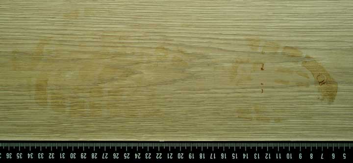
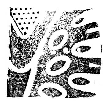
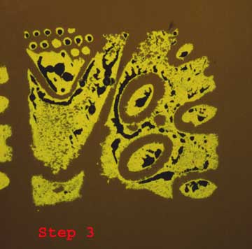
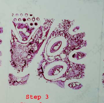
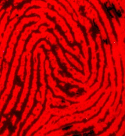
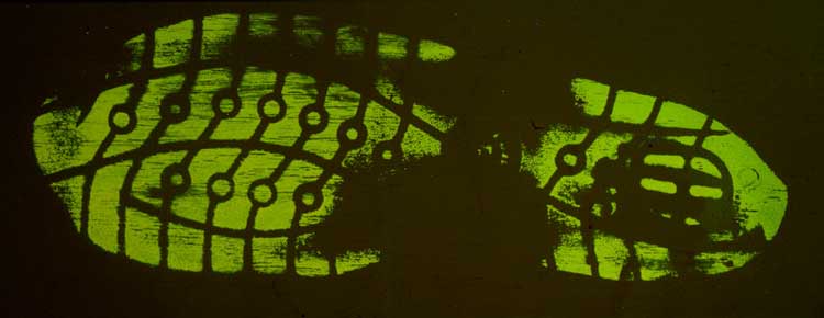
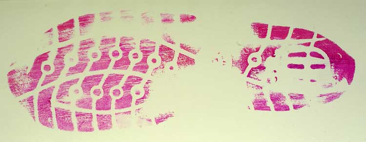
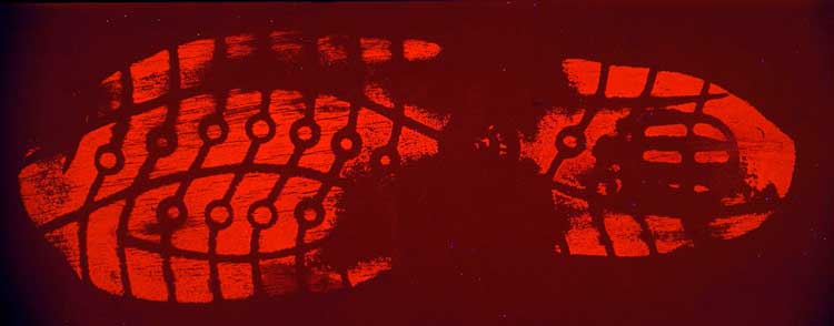
See for example the traces on Photos 1 and 2.
 |
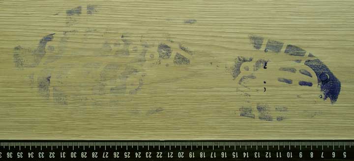 |
I will further discuss the traces shown in Photo 1 and 2 later in this article.
What did not become entirely clear was how one had arrived at the conclusion that LCV is a good enhancement reagent for shoeprints and fingerprints in blood on non-porous surfaces. I suspect that this conclusion was fed by the simplicity using the product.
My questions therefore were:
- In what way did one establish that a blood trace treated with LCV has shown all the latent information that may have been present in a trace?
- Did one subsequently treat the blood trace treated with LVC with another chemical product, in order to verify whether all the latent information in the trace had been developed?
It probably has not been done, as it was not done in the CHEMZIS research: once a blood trace was treated with a chemical reagent or staining solution, the same trace was not used again to look for a possible enhancement with a different reagent or staining solution.
In the past I made this same mistake myself and failed to look for a possible enhancement of a blood trace already treated, even though this was standard procedure with latent fingerprints, using, for instance 5-MTN after having used DFO. I never thought that this would also be possible with blood traces.
So far I have not found any literature in which this follow-up treatment was tested or applied. It did find reports in which it was suggested that in certain cases it would be possible to use a different chemical after LCV, yet no results of a subsequent treatment are published.
As stated earlier, I had a few questions concerning the enhancement of shoeprints and fingerprints placed in blood.
These questions were:
- Can a blood trace that was treated and made visible with Luminol through chemiluminescence be subsequently treated with a blood trace enhancing reagent such as LCV or staining solution such as Acid Yellow 7 or Hungarian Red.
- What is the effect of such a series of treatments to the same trace?
- Is Leuco Crystal Violet really as good for visualising shoeprints and fingerprints in blood as other researchers claim it is and does LCV show latent parts in the trace?
- Do the treatments with the reagents/solutions mentioned under 1 and 3 enhance or compromise the trace?
Mid 2011 I was asked by the forensics department of the Regional Police of Brabant South-East whether I was prepared to share my knowledge with a third-year student of Hogeschool Amsterdam (Amsterdam University of Applied Sciences). This student, Inge van Bijsterveldt, worked there as an intern and intended to do research into the Hungarian Red method.
This presented me with the unique opportunity to find an answer to the questions phrased above, separate from Inge van Bijsterveldt’s own research.
The comparative study I subsequently set up into the enhancement method for shoeprints and fingerprints in blood was performed as follows:
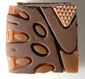
Stamp method
A stamp was made from a piece of sole of about 4 x 4 cm and this was used to make imprints with human blood on a selected surface.
 |
 |
When choosing the stamp method I was aware that when placing the stamp with blood that are so many variables at play that the blood traces can never be identical. Even if the stamping of the blood traces would be automated it still would not be possible to dose the exact same amount of blood every time. For my comparative study, however, such variables are not relevant, because it was focused on the subsequent treatment of the ‘same’ shoeprints and fingerprints, i.e. that one blood trace was subsequently treated with 4 different reagents and staining solutions.
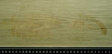
Surface
The surfaces I chose to place the test traces on are all non-porous surfaces:
- Glazed wall tiles, a mottled white/light grey.
- Laminate flooring in pale oak with a wood texture.
Chemicals
The chemical products used were:
- Luminol (according to formulation)
- Leuco Crystal Violet (BVDA)
- Leuco Crystal Violet (according to a local formulation)
- Acid Yellow 7 (BVDA)
- Hungarian Red (BVDA)
- Blood fix: 2% 5-sulphosalicylic acid (BVDA)
(www.bvda.com)
I will not further discuss these products, their effect and composition in this report. My study focuses on the result and the chemicals mentioned have already been extensively described by others.
Blood used
The blood used for this study was human blood including an anticoagulant agent, made available by Inge van Bijsterveldt.
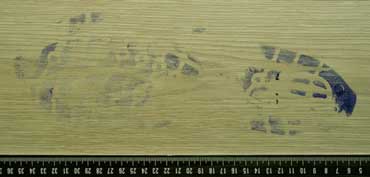
Method of placing the test traces
A small amount of blood was poured into a piece of tissue in a Petri dish, until the tissue became saturated with blood. The stamp was pressed onto the blood-soaked tissue twice, after which 16 consecutive prints were made on a tile, trying to applied similar pressure each and every time a print was made.
This dilution series mimics a track of footsteps where a person first stepped in the blood (indicated as Footstep 1 in image 5) and then walked away from the blood (indicated as Footstep 16). After they were placed, the traces were allowed to dry for about 7 days before they were treated.
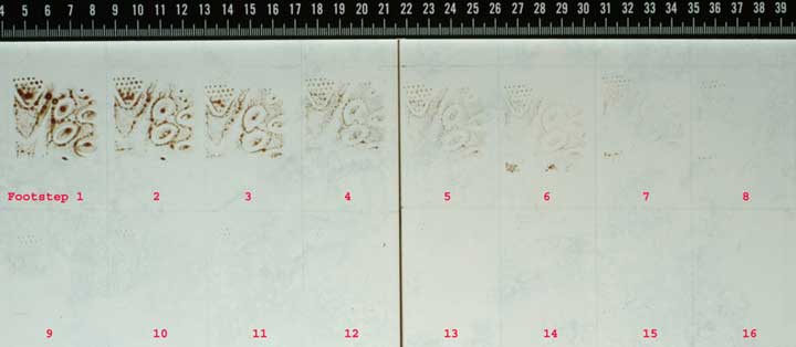 |
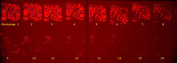
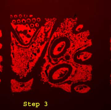
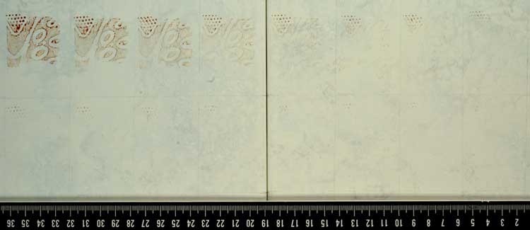
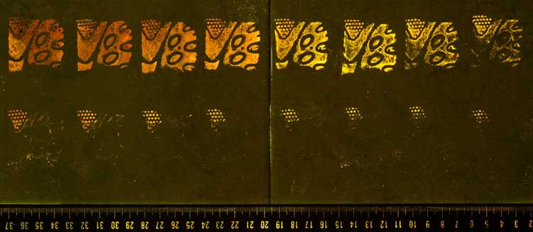
In order to answer my first question, (Can a blood trace that was treated made visible with Luminol through chemiluminescence be subsequently treated with a blood trace enhancing products such as LCV, Acid-Yellow 7 or Hungarian Red?) the blood traces in image no. 5 were first treated with Luminol. In a dark room, the Luminol was nebulised with a high-pressure spraying pistol across the tile with the blood traces three times. With each nebulation chemiluminescence occurred. Throughout the treatment and chemiluminescence the cameras shutter was open (diaphragm: F/6.7, total exposure time: 189 sec. ISO-3200, camera: Canon EOS 300D Digital). Luminol is known for its high sensitivity in reacting with blood, as opposed to certain staining solutions. After the treatment with Luminol the surface clearly showed the reaction with the blood-stained shoe trace stamps. See Photo 6a.
Photographing chemiluminescence at a crime scene is often difficult because it is not certain where the chemiluminescence of a trace will occur. For this reason it is almost impossible to make detail images of shoeprints and fingerprints at an actual crime scene.
The photos taken at a crime scene are better suited to gain an overview of the blood trace pattern at the crime scene. Photographing minute details during the chemiluminescence is all but impossible. The chemiluminescence will disappear after a very short time and once the blood traces have been saturated with Luminol no further chemiluminescence will occur anymore.
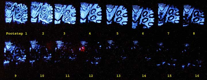 |
Immediately after treatment with Luminol the blood traces were fixed with a 2% solution of 5-sulfosalicylic acid, to prevent running or disruption of the blood traces. It is important to find a chemical reagent that is as similar to Luminol in terms of its sensitivity. This becomes apparent from the extent to which, during treatment with Luminol, the developed test traces formed compared with the pictures.
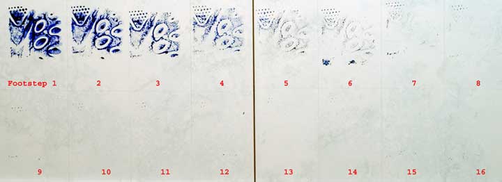
Treatment with LCV
After the Luminol treatment, the same blood traces were treated with LCV, which was allowed 5 minutes to react with the blood. After this, the traces were rinsed with demi water and 3% acetic acid to prevent surface discoloration.
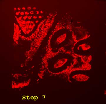
The reaction of LCV in this shoe traces with blood test was mediocre, compared with the picture gained with Luminol in Photo 5. Footstep 7 is hardly even visible after the LCV treatment. What does become clear, though, is that it is possible to apply a chemical treatment to the same blood trace that was treated with Luminol. See Photo 6b for said treatment.
> |
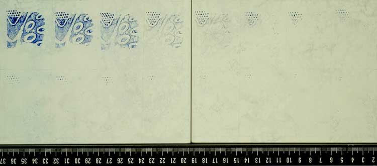
Treatment with Acid Yellow 7
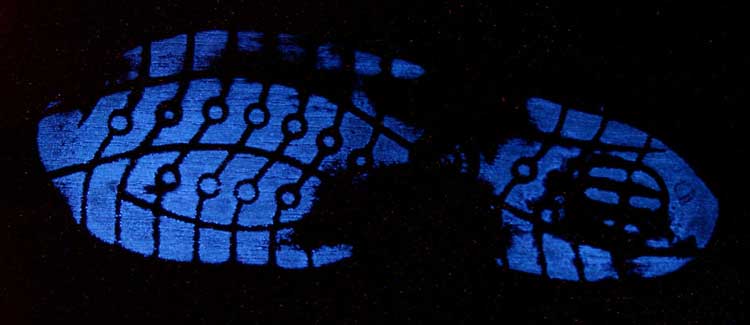
Subsequently, the traces in Photo 6b were treated with Acid Yellow 7. While spraying on the Acid Yellow 7 part of the discoloration caused by the LCV dissolves. One would expect that part of the blood trace would dissolve too, yet this did not happen.
The Acid Yellow 7 was allowed to set for 5 minutes on the blood trace as well, after which it was rinsed with met demi water and 3% acetic acid. The rinsing was thorough, so that no coloring agent of the Acid Yellow 7 was left on the surface, but only on the treated traces. After this the tile was blown dry with compressed air. Photo 7 shows the reaction of Acid Yellow 7 with the blood traces on the surface.
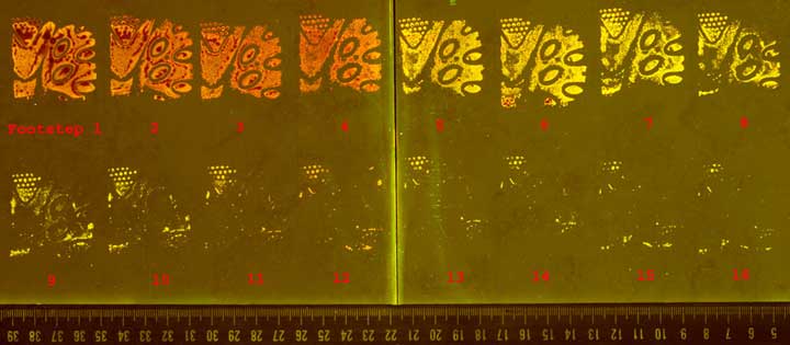 |
Acid Yellow 7 gives a strong fluorescence of the treated blood traces on the surface (Photo 7). The sensitivity of the chemical used is very similar to the sensitivity of Luminol (cf. Photo 5 and 7). The second important benefit, however, is that blood traces treated with this Acid Yellow 7 can be lifted with a white gelatine lifter. This is especially important in case of surfaces that would otherwise discolor, or that show fluorescence themselves, so that the fluorescence of the treated blood trace is not visible on these types of surfaces.
Lifting a trace by means of a gelatine lifter
To achieve good transfer between the trace on the surface and the white gelatine lifter it is advisable to moderately warm the gelatine lifter with a blow dryer, especially if the environment is cold, or if the traces are cold, and to then place the gelatine lifter on the trace without locking in air.
Next a metal or wooden sheet with an approximate weight to 10 kg is placed on the back of the foil. If the surface has some relief it is advised to place a sheet of soft foam rubber between the foil and the pressing sheet so that the white foil remains pressed into the relief. The foil must remain on the trace for at least 30 minutes, although I prefer 60 minutes.
The trace can then be lifted several consecutive times, which may even result in an improvement of the trace. See Photo 8 for the lifted Acid Yellow 7 trace.
 |
Note regarding Hungarian Red
Hungarian Red is a solution made according to a particular formula; the manufacturer (BVDA) always uses the same basic ingredients for this solution, so that, when used for the enhancement of blood traces, the results in terms of sensitivity to blood will always be the same. By Hungarian Red I also refer to a particular method, which is always used in combination with lifting treated traces with a white gelatine lifter. Only when this method has been followed one may refer to the real sensitivity to blood and to other chemical coloring agents in case of Hungarian Red.
In certain studies researchers presented Acid Violet 19 / Acid Fuchsin as Hungarian Red. I should like to want to warm that a non-factory prepared solution with Acid Violet 19 / Acid Fuchsin could yield inferior results compared to the original Hungarian Red as made by BVDA.
As said earlier, I named the method Hungarian Red, and my warning stems from my own experience, because the raw materials used for Acid Violet 19 differ per supplier, even if the chemical formulation and purity is the same.
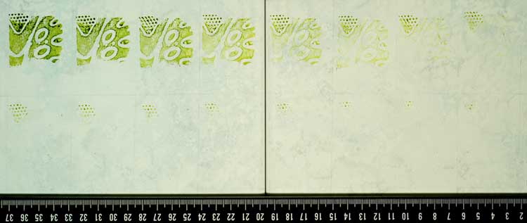
Treatment with Hungarian Red:
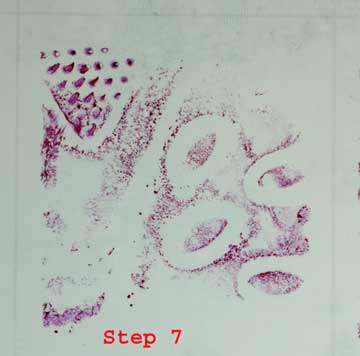
Next the traces were treated once more with the coloring agent Hungarian Red. This was done in the same manner as used for the treatment with Acid Yellow 7. Again this was followed by intense rinsing with demi water/acetic acid. The trace was subsequently dried with compressed air. See Photo 9.
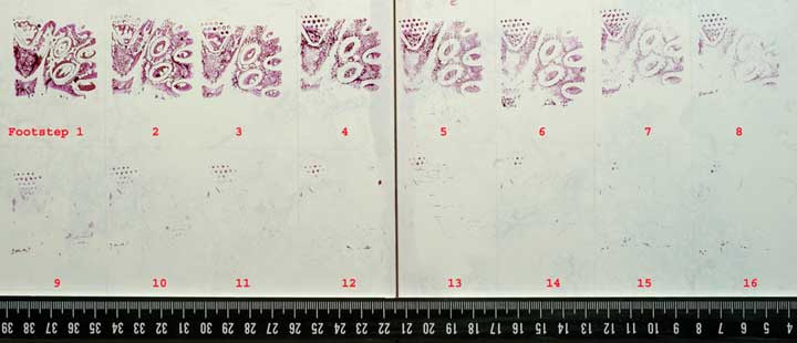 |
Hungarian Red also allows for lifting of the trace with a white gelatine lifter, where the method is identical to lifting Acid Yellow 7. With this method, using Hungarian Red, the sensitivity again is very close to the sensitivity of Luminol.
 |
.
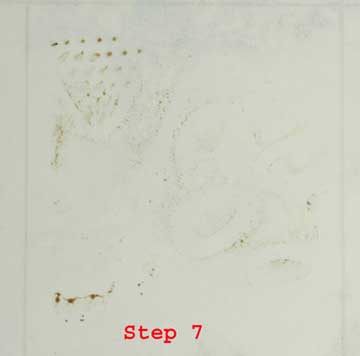
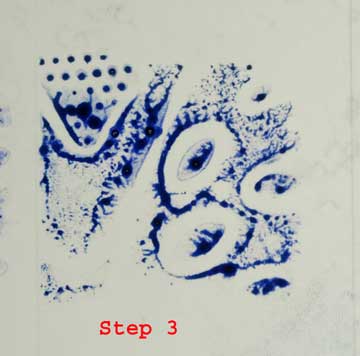
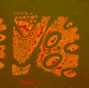
Blow-ups of the above blood traces
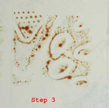
Of the above blood traces blow-ups were made of Foot step 3 and Foot step 7, which allowed for a better assessment of the treatments.
 |
 |
 |
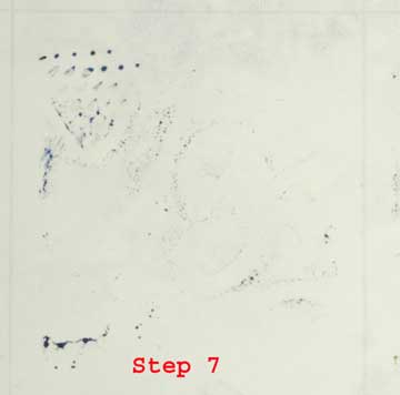 |
No blow-ups were made of the blood traces made visible with Luminol; with the blow-ups shown this was not performed as a first treatment, while as shown above this did in fact take place.
 |
 |
 |
 |
 |
 |
 |
 |
Evaluation of the effect of the subsequent treatment to blood fingerprints
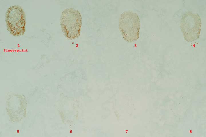
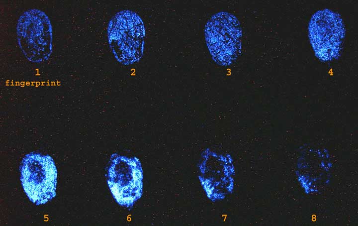
Next, fingerprints were placed on the wall tile (the surface) in the same way. A small amount of blood was poured onto a piece of tissue in a Petri dish, as before, until it the tissue was saturated. One finger was pressed onto the blood-soaked tissue twice, after which 8 consecutive prints were made on the tile, the print being as similar as possible. See Photo 23
 |
 |
Immediately after the Luminol treatment the traces were fixed with a 2% solution of 5-sulfosalicylic acid, which was to prevent any outflow of the blood traces.
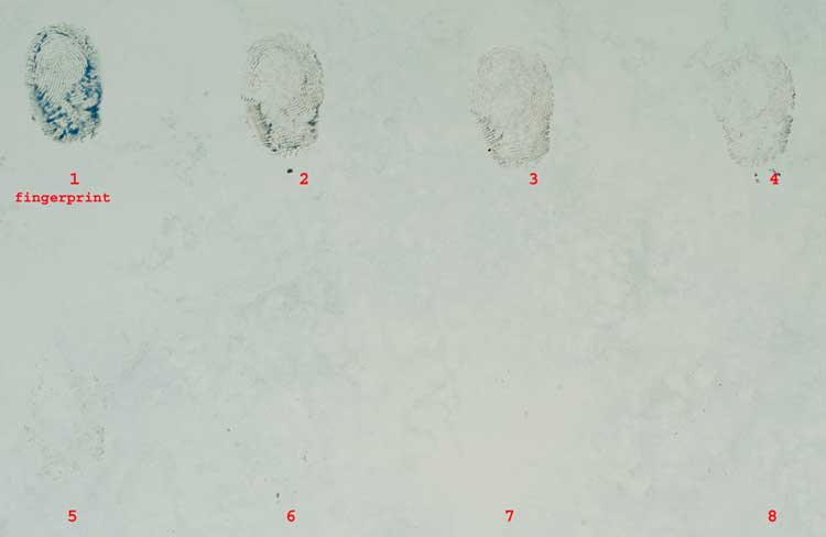
Next, the fingerprints were treated with LCV.
 |
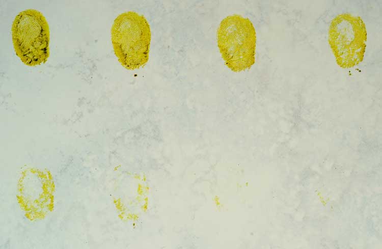
After this treatment the same traces were treated with Acid-Yellow 7. See Photo 26.
 |
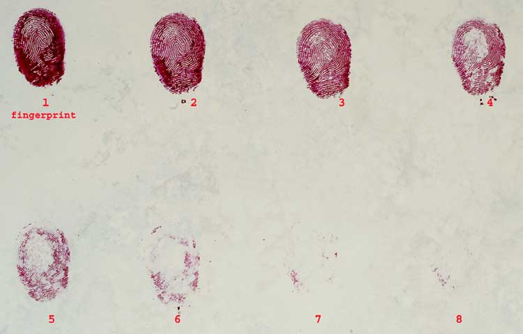
As a final treatment these blood fingerprints received a further treatment with Hungarian Red. See Photo 27.
 |
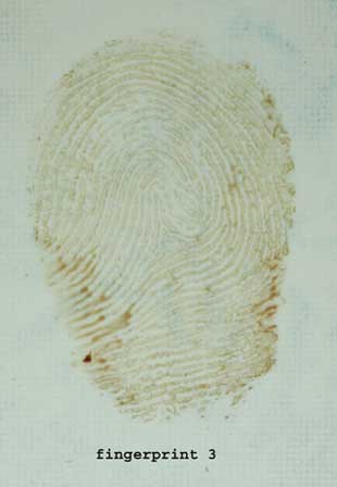
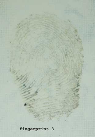
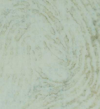
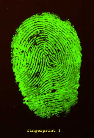
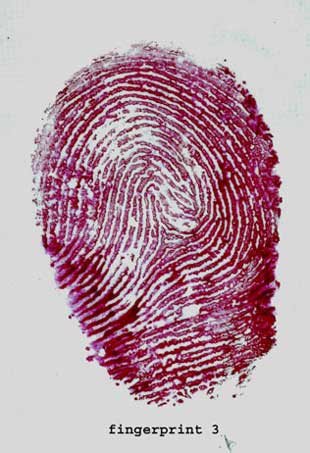
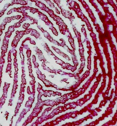
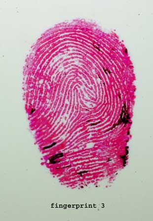
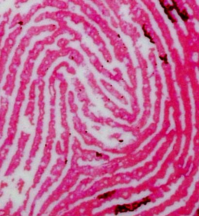
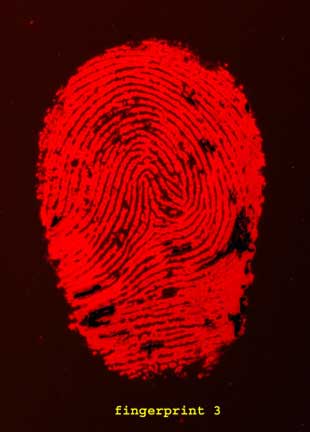
Blow-ups of fingerprint 3
Of blood fingerprint 3 blow-ups were made after each treatment, to allow for a better assessment and comparison. Furthermore, the photo was blown up around the centre of the fingerprint.
 |
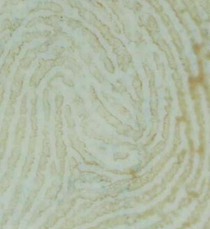 |
When Photos 28 and 29 are compared with Photo 30 (the trace after treatment with LCV) it appear as if the untreated blood trace is better visible in the trace treated with LCV. However, the trace has discolored due to the LCV and this discoloring shows a poorer contrast with the surface.
 |
 |
 |
 |
 |
 |
 |
 |
 |
 |
 |
 |
Webmaster's note: See also the video "Evidence Photography — Basic Concepts"
Sharpness of the different pictures:
Photos taken with chemiluminescence and fluorescence are not as sharp as photos made with white light. This is a normal phenomenon in photography because with chemiluminescence and fluorescence you get diffuse light from the trace.
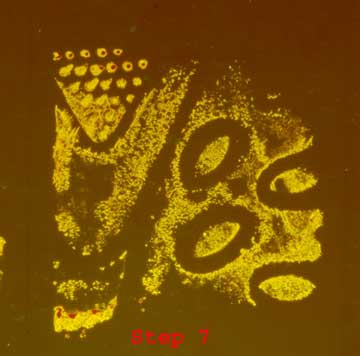
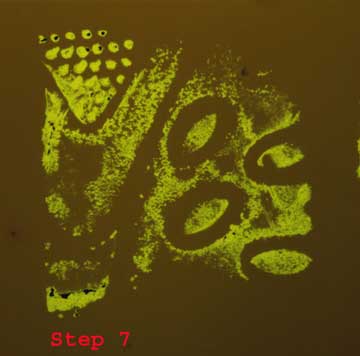
A further look at the shoeprints in photo 1 and 2
Getting back the result of this shoeprint treated with LCV, users of LCV would be pleased with this result and not look on for an improvement of these traces men, as one would suppose that it cannot be made visible any better than this.
The forensic researcher applied this treatment, because this was advised by experts in their reports. As stated above, however, it is possible to retrieve more information from this trace, by subsequently treating it with further products such as Acid Yellow 7 and Hungarian Red.
This also allows for the option of lifting the traces with the white gelatine lifter. By lifting the treated traces the impact (color) of the surface is canceled out entirely, leaving the researcher with a perfect trace.
These are reduced-size pictures of the traces in Photo 1 blood trace and Photo 2 after LCV (page 2). The reduction in size allow for easy comparing.
First this blood shoe trace was treated with Luminol. See Photo 36.
 |
During the Luminol treatment one clearly sees where the blood is present in the trace, both the visible and latent blood. It is this latent blood that one would miss with a LCV treatment alone, meaning that valuable information contained in the trace remains invisible and will be lost.
After the Luminol treatment the LCV treatment as discussed was applied, as is shown in Photo 2. This was followed by the subsequent treatment with Acid Yellow 7, see Photo 37.
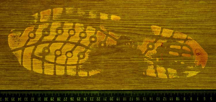 |
Photo 37. Acid Yellow 7 after Luminol and LCV, seen on the surface in fluorescence.
The trace in Photo 37 was then lifted with the white gelatine lifter and photographed in fluorescence.
This clearly shows the added value of lifting, as whatever the color of the surface, one gets to see an excellent trace. See Photo 38.
 |
After treatment the shoe trace underwent subsequent treatment with Hungarian Red and was photographed on the surface with white light. This treatment resulted in a shoe trace with a sharper contrast with the surface. See Photo 39. Note that if the surface would be such that the treated blood trace would not or hardly be visible, e.g. because it was black, this has no impact whatsoever on the end result with, because these two staining solutions can be lifted with a white gelatine lifter.
 |
 |
 |
Note regarding Luminol and LCV
Both Luminol and LCV are peroxidase reagents. One would therefore expect that LCV would no longer chemically react with the blood traces after they have been treated with Luminol. This could then be the reason why LCV shows a bad results.
However, during the forementioned experiments of Inge van Bijsterveldt, it appeared that a direct chemical reaction (without being treated with Luminol first) of LCV with the blood traces resulted in the same poor results. These are the results Inge van Bijsterveld obtained:
 |
 |
 |
 |
 |
Conclusion
- I don't think a clearer example of consecutive treatments of shoeprints and fingerprints placed in blood can be given.
- Visible traces;
- Partially visible traces;
- Latent traces;
- Traces that are a combination of A, B and C.
- With Luminol the traces A, B, C and D mentioned above were clearly visible.
- In selecting a blood enhancement product it is important to select a chemical that is similar to Luminol in terms of its sensitivity to blood and to select one that will also show latent traces to the greatest possible extent.
- In this study it became clear that the treatment of blood traces with Luminol does not have a negative effect on the shoeprints and fingerprints seen as shape traces.
- Subsequently, these traces could be further treated with LCV or a staining solution such as Acid Yellow or Hungarian Red with an enhanced result compared with LCV.
- The product Leuco Crystal Violet only enhances the contrast of visible blood traces, but it does not show latent traces. LCV did not have the same sensitivity as Luminol. LCV is not suited as a replacement of Luminol for detecting latent blood traces.
- Leuco Crystal Violet is a chemical reagent that in this study proved unsuited for making visible shoeprints and fingerprints in blood on NON-POROUS surface materials.
- If LCV would be used in NON-POROUS materials to enhance the contrast of visible blood traces there always has to be a subsequent treatment because it has not yet been established whether the trace contains any latent parts that were not shown with LCV. The chemical chosen for this, depending on the intended effect, is either Acid Yellow 7 or Hungarian Red.
- To achieve an optimum result with Acid Yellow 7 and/or Hungarian Red one must always lift the shoeprint and fingerprints made visible with these staining solutions using a white gelatine lifter. The special property of the white gelatine lifter is that it absorbs coloring agents and makes them fluorescent. Hungarian Red will only show fluorescence if the trace has been lifted with the white gelatine lifter and it will not fluoresce on a surface. A second property of the white gelatine lifter is that it does not fluoresce itself so that the lifted trace gets an even sharper contract. By lifting the blood traces treated with these staining solutions the impact of the surface (both color and texture) is cancelled out entirely. Thus, the trace is copied from the surface which results in a noise-free image of the trace, while the trace itself on the surface is not compromised.
- The gelatine lifter should be left under a weight of approx. 10 kg for at least 30 minutes to ensure a proper transfer of the trace to the gelatine lifter. Next the lifted traces without the “protective layer” must be photographed within 60 minutes, with white light and in fluorescence with light of the appropriate wave length.
- Even though in this study I did not examine the effect of LCV on surfaces that are porous, I think we may assume that even with these materials one must be very cautious with suspected latent blood traces.
- In this study two different solutions of LCV were used in a large number of test traces, yet the results for the latent traces was the same for both LCV solutions.
- Conclusions about LCV drawn by authors/researchers in the past may have been correct given the knowledge available to them at the time, yet in 2012 they have been superseded by science.
- My study was not intended to suggest that a series of subsequent treatments should always be performed with one and the same trace the crime scene. Even the use of Luminol is not necessary in all cases, if the traces at the crime scene already are already clear about this.
- What is important to note, though, is that after the use of Acid Yellow 7 the chemical Hungarian Red can be used to achieve a different effect with the trace and the surface and that the quality of the subsequent treatment will not deteriorate.
- When choosing an excellent quality staining solution such as Acid Yellow 7 or Hungarian Red in combination with lifting the traces by means of a gelatine lifter, one can be certain that 99% of all information present in the trace is retrieved. This is demonstrated by the comparison of the photos of the traces and those of the Luminol treatment.
- Acid Yellow 7 and Hungarian treatment come close to the sensitivity to blood of Luminol.
- As for the quality of the treated shoeprints and fingerprints after the chemical treatments the conclusion is very clear: the quality of the traces was not compromised by the subsequent treatments.
- This is best demonstrated by the fingerprints in blood, which after all treatments show an excellent papillary line print with pores and clear edges, so that these dactyloscopic traces are even eminently suited for an examination according to the standards and views of Ridgeology.
Shoe and finger traces placed in blood can be subdivided into:
Method of treatment
- Photograph the trace.
- Fix the trace with 2% 5-sulphosalicylic acid, leave to set for 5 minutes.
- Treat the trace with Acid Yellow 7 or Hungarian Red, leave to set for 5 minutes.
- Rinse the trace profoundly with acetic acid/demi water ( 30:1000 )
- Immediately blow the trace dry with compressed air.
- Check is the trace is dry.
- Photograph the trace with white light and then, if needed, in fluorescent, wave length to be used depending on the chemical product used, either 450 nm. or 530 nm. Then observe with yellow, orange or red filter, and photograph with a similar filter.
- Lift the trace with fresh white gelatine lifter (BVDA) by applying the gelatine lifter without locked-in air to the dry trace and applying pressure (according to the method described earlier in this report).
- Leave the gelatine lifter on the trace for at least 30 minutes, then lift the trace and immediately photograph it with white light and then in fluorescence with an appropriate wavelength. It is crucial to make photos of the lifted material within one hour after lifting, as otherwise there is a risk that the trace on the gelatine lifter will flow and that it will lose its sharpness.
- If necessary, lift the trace a second time, which may give better results compared with the first lift.
- Next, if necessary, the trace, after being treated with another staining solution, depending on the first treatment selected, can be treated once more, for which the same method is followed as above, minus step 2.
Cameras used:
For this study, two cameras were used:
- Canon EOS 300D.
- Nikon D 100.
- The filters used Yellow filter and bandpass filter 610/30 nm.
- Excitation wave length for Acid Yellow: 450 nm
- Excitation wave length for Hungarian Red: 530 nm
Literature
Bossers, L.C.A.M., Roux, C., Bell. M., Mcdonagh, A.M. (2011). Methods for the enhancement of fingermarks in blood. Forensic Science International, 210, 1-11.
Farrugia, K.J., Savage, K.A., Bandey, H., NicDaéid, N. (2010). Chemical enhancement of footwear impressions in blood on fabric – part 1: Protein stains. Science and Justice, 51, 99-109.
Farrugia, K.J., Savage, K.A., Bandey, H., Ciuksza, T., NicDaéid, N. (2010). Chemical enhancement of footwear impressions in blood on fabric – part 2: Peroxidase reagents. Science and Justice, 51, 110-121
Bodziak, W.J. (1995). Use of leuco crystal violet to enhance shoe prints in blood. Forensic Science International, 82, 45-52.
Naber, W.M., Theeuwen, A.B.E., Limborgh, J., Keereweer, I., Velders, T., Drok, J.W., Barneveld, S.V. (1996). CHEMZIS, Chemische methoden voor het zichtbaar maken en/of verbeteren van schoensporen gezet met bloed.
M.J.M. Velders, Fluorescing traces in blood on white gelatine lifters with Hungarian red, 81st Educational Conference of the International Association of Identification 1996, Greensboro, NC.
Theeuwen, A.B.E., Limborgh, J., Keereweer, I., Naber, W.M, Velders, T., Drok, J.W., Barneveld, S.V. (1998) Enhancement of Footwear Impressions in blood, Forensic Science International 95 (2) (1998) 133- 151
Acknowledgement
I would like to thank intern Inge van Bijsterveldt for providing me with the blood samples and for her assistance with the Luminol procedure and photo 42 – 46. Data and photos in this report may only be used with a reference to the source and the name of the author. Theo Velders (email: theovelders2@live.nl).
 New Insight into the Chemical Improvement of Shoeprints and Fingerprints Placed with Blood on Non-Porous Surfaces Copyright: © 2012 by Theo Velders. Copyright for this article is retained by the author, with publication rights granted to the Crime Scene Investigation Network. This is an Open Access article distributed under the terms of the Creative Commons Attribution-NonCommercial-NoDerivatives 4.0 International License which permits unrestricted noncommercial use, distribution, and reproduction, provided the original work is properly cited and not changed in any way. Based on a work at https://www.crime-scene-investigator.net/new-insight-into-the-chemical-improvement-of-shoeprints-and-fingerprints-placed-with-blood-on-non-porous-surfaces.html.
Article submitted by the author. The Crime Scene Investigator Network gratefully acknowledges the author for allowing us to reproduce the article .
Article posted November 28, 2019